When Seconds Count
July 2016
Strokes.
They happen when the blood that is circulating within the brain is cut off or reduced in an area resulting in a loss of oxygen to that part of the brain. If not treated right away the brain can die.
Thanks to nuclear medicine, doctors can better assess and treat stroke patients, saving lives.
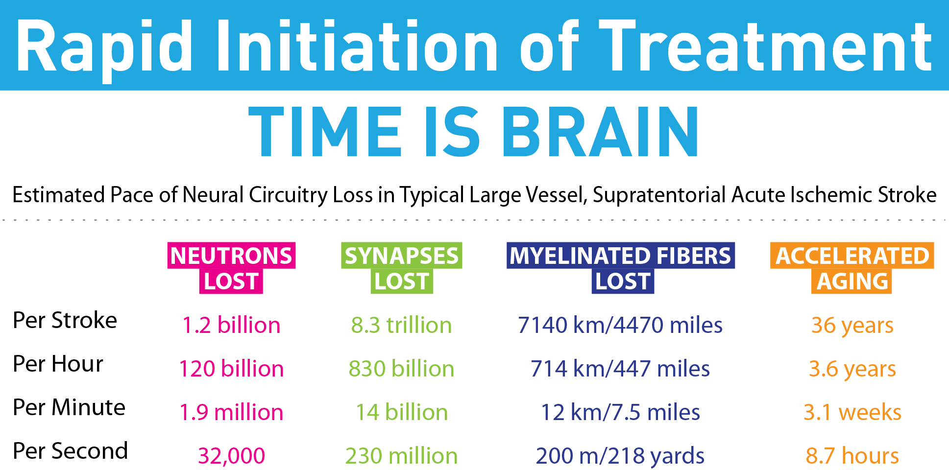
With a stroke, every second counts. Neurons or nerve cells are lost at a rate of 1.9 million per minute when the brain is deprived of oxygen. The loss of these and other key brain transmitters, or synapses leads to accelerated aging in the brain.

(Chart Courtesy of Dr. Timo Krings, Head of Neuroradiology, Joint Department of Medical Imaging, Toronto Western Hospital & University Health Network)
Dramatic treatment is required to save a patient’s life, everything has to move rapidly.
“2:00am: I was called for a stroke at the hospital. By 2:20 the team was at the hospital and by 2:40 we had removed the blood clot and the patient was off the operating table,” states Dr. Timo Krings, Head of Neuroradiology, University Health Network, Toronto Western Hospital.
From initial imaging to opening the blocked blood vessel and restoring blood flow happens in just a matter of minutes.
The proper diagnosis and successful treatment of strokes is thanks to a branch of Radiology known as Interventional Neuroradiology.
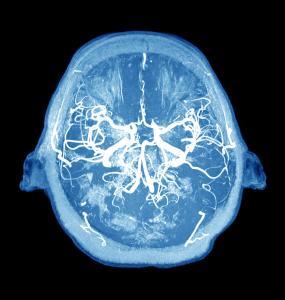
Interventional radiology involves a uses modern Nuclear Magnetic Resonance Imaging or Computed Tomography to identify where the blockage is within the brain and its size.
As Dr. Krings points out, imaging plays a major role in proper diagnosis and treatment.
“The imaging part is just as important because if we choose the wrong patient we can make things worse,” he explains. “If we open a vessel that was occluded for too long, l the dead brain won’t recover but the patient can be in danger because blood will rush to the dead brain, which can lead to a hemorrhage. Therefore the correct identification of right patients is as important as the treatment itself.”
Once the scan identifies a suitable patient, the treatment can begin.
Interventional Neuroradiology involves a minimally invasive procedure whereby an artery is punctured and small tubes known as catheters are placed into the blood vessels. In these small tubes, doctors can push in stents or devices into the blood vessels in order to treat the blood vessel from the inside out.
 Symptoms you may be having a stroke: FAST
Face: Drooping eyelid or numbness on one side of your face. Vertigo or a whirling loss of balance
Arm: Unable to move your hand or arm on one side of your body
Speech: You can’t speak properly or speech is garbled
Time: Time is of essence and you should telephone 9-1-1 , if you think you may be having a stroke call 9-1-1
Dr. Krings points to a chain of stroke awareness and treatment. Beginning with the patient to be able to identify the symptoms of a stroke to emergency medical service (EMS) workers who can rapidly identify and take patients to stroke centers where emergency room (ER) teams can fast track patients into imaging.
“It’s a team effort between the patient, EMS, the ER doctor, neurologist, radiologist, dietician and rehab specialists. All of them have to play together to get the best outcome,” says Krings.
New Canadian guidelines call for the two pronged technique of imaging and interventional radiology as the best life-saving method to for the diagnosis and treatment of strokes.


