Nuclear Imaging
Radiation from radioisotopes passes through people’s bodies, which is how X-rays work. When taking an X-ray, the radiation starts on one side of the person’s body and passes through the body to film on the other side. Nuclear imaging is similar.
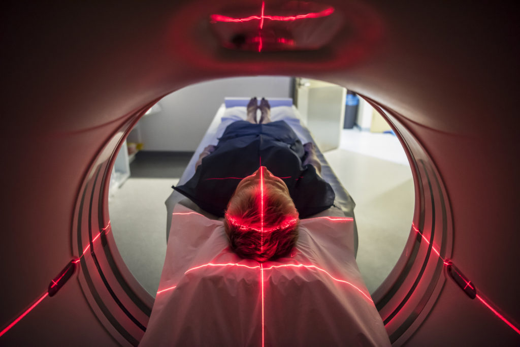
However, health-care professionals inject isotopes into the patient’s body. The radiation is emitted and captured on film. This material, often referred to as a tracer, emits gamma radiation from inside the patient and is detected by an imaging device, which the patient sits in or near.
Canadian nuclear medical professionals (radiologists, technologists, nuclear pharmacists and nuclear physicists) have spent decades investigating different medical isotopes. This research has led to more effective nuclear treatments. Body tissues, such as bones and blood, absorb various isotopes at different rates, which means a medical team can image a specific area while limiting radiation exposure to the rest of the body.
Tracing techniques can also assess bone growth or areas of restricted blood flow, and produce time-lapse functional images of organs, such as the brain, heart, liver, lungs and kidneys.
Continued advances in research enhance the precision of life-saving nuclear diagnostic procedures while minimizing radiation exposure. According to the Health Physics Society, standard nuclear medicine procedures involve doses between 1.8 and 14.1 millisieverts or between 0.6 and 4.7 times the dose received annually from natural background radiation in most parts of Canada. These doses, while higher than for X-rays, are considered very safe, especially as they provide life-saving information.